 Intestinal Tuberculosis Intestinal Tuberculosis is a TB disease of the intestine. Tuberculosis is primarily a Lung Infection, but it can infect other areas of the body as well. Intestinal Tuberculosis frequently complicates Lung Infections with Tuberculosis. In addition, milk, which contains tuberculi bacteria, may also infect the intestine. Symptoms of the disease include fever, anorexia, nausea, flatulence (gas), food intolerance, abdominal cramps in lower right abdomen, abdomen distention after eating, and weight loss. Sometimes Intestinal TB may manifest no symptoms. [External Links]Information from ecureme.comInformation from eMedicine (Radiology)
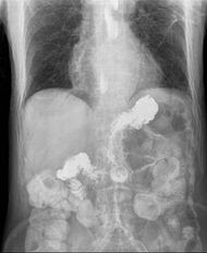 Gastroesophageal reflux disease Gastroesophageal reflux disease (GERD), Gastro-oesophageal reflux disease (GORD), Gastric reflux disease, or Acid reflux disease is defined as chronic symptoms or mucosal damage produced by the abnormal reflux in the esophagus. This is commonly due to transient or permanent changes in the barrier between the esophagus and the stomach. This can be due to incompetence of the lower esophageal sphincter, transient lower esophageal sphincter relaxation, impaired expulsion of gastric reflux from the esophagus, or a hiatus hernia. If the reflux reaches the throat, it is called laryngopharyngeal reflux disease. The most-common symptoms of GERD are heartburn, regurgitation, and trouble swallowing (dysphagia). The less-common symptoms include pain with swallowing (odynophagia), excessive salivation (this is common during heartburn, as saliva is generally slightly basic and is the body's natural response to heartburn, acting similarly to an antacid), nausea, and chest pain. GERD sometimes causes injury of the esophagus. These injuries may include Reflux esophagitis—necrosis of esophageal epithelium causing ulcers near the junction of the stomach and esophagus, Esophageal strictures — the persistent narrowing of the esophagus caused by reflux-induced inflammation, Barrett's esophagus—metaplasia (changes of the epithelial cells from squamous to columnar epithelium) of the distal esophagus, and Esophageal adenocarcinoma — a rare form of cancer. Several other atypical symptoms are associated with GERD, but there is good evidence for causation only when they are accompanied by esophageal injury. These symptoms are chronic cough, laryngitis (hoarseness, throat clearing), asthma, erosion of dental enamel, dentine hypersensitivity, and damaged teeth. GERD may be difficult to detect in infants and children. Symptoms may vary from typical adult symptoms. GERD in children may cause repeated vomiting, effortless spitting up, coughing, and other respiratory problems. Inconsolable crying, failure to gain adequate weight, refusing food, bad breath, and belching or burping are also common. Children may have one symptom or many — no single symptom is universal in all children with GERD. Common symptoms of Pediatric Reflux are irritability and pain, sometimes screaming suddenly when asleep. They may also have constant or sudden crying or "colic" like symptoms. Babies can be inconsolable especially when laid down flat. They may have poor sleep habits typically with arching their necks and back during or after feeding. They may have excessive possetting or vomiting, frequent burping or frequent hiccups, excessive dribbling or runny nose, swallowing problems, gagging and choking. Babies are often very gassy and extremely difficult to "burp" after feeds, they may refuse feeds or may frequent feeds for comfort. They may also manifest night time coughing. Extreme cases of acid reflux can cause apnea and respiratory problems such as asthma, bronchitis and pneumonia if stomach contents are inhaled. They may also have bad breath – smelling acidy, and rancid/acid smelling diapers with loose stool. Bowel movements can be very frequent or babies can be constipated. [External Links]More information from medicinenet.comMore information from eMedicineExtra information from gicare.com
Acute Gastroenteritis comes from the word Acute, which means sudden, and Gastroenteritis, a disease of the Gastrointestinal Tract characterized by inflammation or irritability. Acute gastroenteritis could be more simply called a long, and potentially lethal bout of stomach flu. The most common symptoms are diarrhea, vomiting and stomach pain, because whatever causes the condition inflames the gastrointestinal tract. Acute gastroenteritis is quite common among children, though it is certainly possible for adults to suffer from it as well. While most cases of gastroenteritis last a few days, acute gastroenteritis can last for weeks and months. Numerous things may cause acute gastroenteritis. Bacterial infection is frequently a factor, and infection by parasites like giardia can cause acute gastroenteritis to last for several weeks. Viruses can also cause lengthy stomach flu, particularly rotaviruses and noroviruses. Accidental poisoning or exposure to toxins may also instigate acute gastroenteritis as well. When a person does not recover from stomach flu symptoms within a day or so, it is usually a good idea to see a doctor. Some types of acute gastroenteritis will not resolve without antibiotic treatment, especially when bacteria or exposure to parasites are the cause. Physicians may want to diagnose the cause by analyzing a stool sample, when stomach symptoms remain problematic. Another reason to seek medical treatment is that some forms of acute gastroenteritis mimic appendicitis, which may require emergency treatment. As well, young children run an especially high risk of becoming dehydrated during a long course of the stomach flu. One should receive directions regarding how to help affected kids or adults get more fluids. Sometimes children, those with compromised immune systems, and the elderly may require hospitalization and intravenous fluids. Dehydration can actually cause greater nausea, and can begin to cause organ shut down if not properly addressed. Even through causes for acute gastroenteritis vary, methods of transmission from one person to another usually remain the same. Generally, contact with the fecal matter of a person with the condition and then improperly washing or not washing the hands causes acute gastroenteritis to be quite contagious. Proper hand washing for both the ill person, and well people in the family is always encouraged. Other methods of transmission of acute gastroenteritis can include eating food or drinking liquids contaminated with bacteria or parasites. For example, poorly cooked hamburger might result in a very severe case of acute gastroenteritis due to exposure to E. coli, a sometimes lethal bacterial infection in young children. Drinking improperly treated water, or drinking from streams and lakes can expose one to giardia, which can leave one ill for many weeks, without treatment. [External Links]Related link from eMedicine
 Gastroenteritis Gastroenteritis (also known as gastro, gastric flu, tummy bug in the United Kingdom, and stomach flu, although unrelated to influenza) is inflammation of the gastrointestinal tract, involving both the stomach and the small intestine and resulting in acute diarrhea. The inflammation is caused most often by infection with certain viruses, less often by bacteria or their toxins, parasites, or adverse reaction to something in the diet or medication. At least 50% of cases of gastroenteritis as foodborne illness are due to norovirus. Another 20% of cases, and the majority of severe cases in children, are due to rotavirus. Other significant viral agents include adenovirus and astrovirus. Different species of bacteria can cause gastroenteritis, including Salmonella, Shigella, Staphylococcus, Campylobacter jejuni, Clostridium, Escherichia coli, Yersinia, and others. Some sources of the infection are improperly prepared food, reheated meat dishes, seafood, dairy, and bakery products. Each organism causes slightly different symptoms but all result in diarrhea. Colitis, inflammation of the large intestine, may also be present. Risk factors include consumption of improperly prepared foods or contaminated water and travel or residence in areas of poor sanitation. It is also common for river swimmers to become infected during times of rain as a result of contaminated runoff water. The incidence is 1 in 1,000 people. Gastroenteritis can be classified as either viral or bacterial. Gastroenteritis often involves stomach pain or spasms (sometimes to the point of being incapacitated), diarrhea and/or vomiting, with noninflammatory infection of the upper small bowel, or inflammatory infections of the colon. It usually is of acute onset, normally lasting 1-6 days, in some cases even longer and self-limiting. Among the signs and symptoms are Nausea and vomiting, Diarrhea, Loss of appetite, Abdominal pain, Abdominal cramps, Bloody stools (dysentery - suggesting infection by amoeba, Campylobacter, Salmonella, Shigella or some pathogenic strains of Escherichia coli), and Fainting and Weakness. The main contributing factors include poor feeding in infants. Diarrhea is common, and may be followed by vomiting. Viral diarrhea usually causes frequent watery stools, whereas blood stained diarrhea may be indicative of bacterial colitis. In some cases, even when the stomach is empty, bile can be vomited up. A child with gastroenteritis may be lethargic, suffer lack of sleep, run a low fever, have signs of dehydration (which include dry mucous membranes), tachycardia, reduced skin turgor, skin color discoloration, sunken fontanelles, sunken eyeballs, darkened eye circles, glassy eyes, poor perfusion and ultimately shock. Symptoms occur for up to 6 days on average. Given appropriate treatment, bowel movements will return to normal within a week after that. [External Links]More information from MedlinePlusComplete information from emedicinehealth.comMore Information from medicinenet.com
Aase syndrome or Aase-Smith syndrome is a rare inherited disorder characterized by anemia with some joint and skeletal deformities. The anemia is caused by underdevelopment of the bone marrow, which is where blood cells are formed. It is named after the American pediatricians Jon Morton Aase and David Weyhe Smith. Some of the symptoms it manifests are mildly slowed growth, pale skin, delayed closure of fontanelles (soft spots), narrow shoulders, triple jointed thumbs, absent or small knuckles, decreased skin creases at finger joints, inability to fully extend the joints from birth (congenital contractures), cleft palate, deformed ears, droopy eye lids. [External Links]Information complete with diagnosis and more from wrongdiagnosis.comInformation from umm.edu
Colic or Abdominal cramps are caused by involuntary muscle contractions that can be very painful. Cramps or pain may occur when any of the muscles in the abdomen or in the walls of the bowels are stretched or strained. Because many internal organs are located in this region, it is hard to know the exact cause of cramping without further investigation. Cramps in the abdomen can occur from: food moving through the stomach and intestines, mild food poisoning that is usually accompanied by diarrhea, stress and anxiety, gastritis (an inflammation of the stomach lining), menstruation, disease or infection in the uterus, and pregnancy, childbirth, and changing hormone levels during breast-feeding. Symptoms such as rumbling and gurgling noises, belching, passing gas, nausea, or vomiting may be felt. Diarrhea or constipation may occur. Urinary problems or loss of hunger may also be present. More serious conditions, such as appendicitis or gall bladder attacks, will usually cause severe and constant cramping. Cramping that last more than one-half hour, and are not related to menstruation or a known digestive condition (such as colitis), requires medical attention. [External Links]Information from uihealthcare.comInformation from wrongdiagnosis.comInformation from MedlinePlus
A steroid hydroxylase system found in adrenal tissues. It catalyzes the 11-hydroxylation of steroids in the presence of molecular oxygen and consists of an NADPH-specific flavoprotein, a non-heme iron protein, and cytochrome P-450. Its deficiency is a cause of congenital adrenal hyperplasia. [External Links]Information from gpnotebook.co.ukInformation from OMIM
 An aneurysm (or aneurism) is a localized, blood-filled dilation (balloon-like bulge) of a blood vessel caused by disease or weakening of the vessel wall. Aneurysms most commonly occur in arteries at the base of the brain (the circle of Willis) and in the aorta (the main artery coming out of the heart, a so-called aortic aneurysm). As the size of an aneurysm increases, there is an increased risk of rupture, which can result in severe hemorrhage or other complications including sudden death. Diagnosis of a ruptured cerebral aneurysm is commonly made by finding signs of subarachnoid hemorrhage on a CT scan (Computed Tomography, sometimes called a CAT scan, a computerized test that rapidly X-rays the body in cross-sections, or slices, as the body is moved through a large, circular machine). If the CT scan is negative but a ruptured aneurysm is still suspected, a lumbar puncture is performed to detect blood in the cerebrospinal fluid (CSF). Computed Tomography Angiography (CTA) is an alternative to the traditional method and can be performed without the need for arterial catheterization. This test combines a regular CT scan with a contrast dye injected into a vein. Once the dye is injected into a vein, it travels to the brain arteries, and images are created using a CT scan. These images show exactly how blood flows into the brain arteries. [External Links]Information from nhlbi.nih.govInformation from MedlinePlusInformation from mercksource.com
 The 1918 flu pandemic (commonly referred to as the Spanish flu) was an influenza pandemic that spread to nearly every part of the world. It was caused by an unusually severe and deadly Influenza A virus strain of subtype H1N1. Historical and epidemiologic data are inadequate to identify the geographic origin of the virus. Most of its victims were healthy young adults, in contrast to most influenza outbreaks which predominantly affect juvenile, elderly, or otherwise weakened patients. The pandemic lasted from March 1918 to June 1920, spreading even to the Arctic and remote Pacific islands. It is estimated that anywhere from 20 to 100 million people were killed worldwide, or the approximate equivalent of one third of the population of Europe, more than double the number killed in World War I. This extraordinary toll resulted from the extremely high illness rate of up to 50% and the extreme severity of the symptoms, suspected to be caused by cytokine storms. The disease was first observed at Fort Riley, Kansas, United States, on March 4, 1918, and Queens, New York, on March 11, 1918. In August 1918, a more virulent strain appeared simultaneously in Brest, France, in Freetown, Sierra Leone, and in the U.S. at Boston, Massachusetts. The Allies of World War I came to call it the Spanish flu, primarily because the pandemic received greater press attention after it moved from France to Spain in November 1918. Spain was not involved in the war and had not imposed wartime censorship. Scientists have used tissue samples from frozen victims to reproduce the virus for study. Given the strain's extreme virulence there has been controversy regarding the wisdom of such research. Among the conclusions of this research is that the virus kills via a cytokine storm, which explains its unusually severe nature and the unusual age profile of its victims (the virus caused an overreaction of the body's immune system—the strong immune systems of young adults ravaged the body, while the weaker immune systems of children and middle-aged adults caused fewer deaths). [External Links]Information from virus.stanford.eduLink to March 21, 1997 news regarding Spanish Flu
 Scrofula Scrofula is any of a variety of skin diseases; in particular, a form of tuberculosis, affecting the lymph nodes of the neck. It is often informally or historically known as 'King's Evil', referring to the method of treatment many sufferers used, in some cases in England up to the reign of King Charles II. In adults it is caused by Mycobacterium tuberculosis and in children by nontuberculous mycobacteria. The word comes from the Latin scrofula, meaning brood sow. Scrofula is the term used for tuberculosis of the neck, or, more precisely, a cervical tuberculous lymphadenopathy. Scrofula is usually a result of an infection in the lymph nodes, known as lymphadenitis and is most often observed in immunocompromised patients (about 50% of cervical tuberculous lymphadenopathy). About 95% of the scrofula cases in adults are caused by Mycobacterium tuberculosis, but only 8% of cases in children. The rest are caused by atypical mycobacterium ( Mycobacterium scrofulaceum) or nontuberculous mycobacterium (NTM). The most usual signs and symptoms are the appearance of a chronic, painless mass in the neck, which is persistent and usually grows with time. The mass is referred to as a "cold abscess", because there is no accompanying local color or warmth and the overlying skin acquires a violaceous (bluish-purple) color. NTM infections do not show other notable constitutional symptoms, but scrofula caused by tuberculosis is usually accompanied by other symptoms of the disease, such as fever, chills, malaise and weight loss in about 43% of the patients. As the lesion progresses, skin becomes adhered to the mass and may rupture, forming a sinus and an open wound. Diagnosis is usually performed by needle aspiration biopsy or excisional biopsy of the mass and the histological demonstration of stainable acid-fast bacteria in the case of infection by M. tuberculosis (Ziehl-Neelsen stain), or the culture of NTM using specific growth and staining techniques. [External Links]Information from eMedicineInformation from umm.eduInformation from MedlinePlus
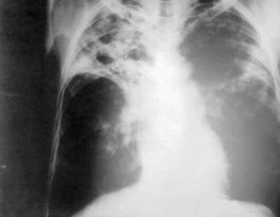 Pulmonary tuberculosis Pulmonary tuberculosis (TB) is a contagious bacterial infection that mainly involves the lungs, but may spread to other organs. Pulmonary tuberculosis is caused by the bacteria Mycobacterium tuberculosis (M. tuberculosis). You can get tuberculosis by breathing in air droplets from a cough or sneeze of an infected person. Most people will recover from primary TB infection without further evidence of the disease. The infection may stay asleep or nonactive (dormant) for years and then reactivate. Most people who develop symptoms of a TB infection first became infected in the past. However, in some cases, the disease may become active within weeks after the primary infection. The following people are at higher risk for active TB: Elderly, Infants, People with weakened immune systems, for example due to AIDS, chemotherapy, or antirejection medicines given after an organ transplant. The risk of contracting TB increases if you: Are in frequent contact with people who have the disease, Have poor nutrition, Live in crowded or unsanitary living conditions. The following factors may increase the rate of TB infection in a population: Increase in HIV infections, Increase in number of homeless people (poor environment and nutrition), The appearance of drug-resistant strains of TB. The primary stage of the disease usually doesn't have symptoms. When symptoms do occur, they may include: Cough (sometimes producing phlegm), Coughing up blood, Excessive sweating, especially at night, Fatigue, Fever, and Unintentional weight loss. Other symptoms that may occur with this disease are breathing difficulty, chest pain, and wheezing. [External Links]Complete information from umm.eduInformation from healthguide.howstuffworks.comMore information from eMedicine
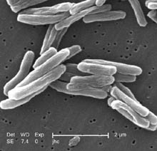 Tuberculosis Tuberculosis (abbreviated as TB for tubercle bacillus or Tu berculosis) is a common and often deadly infectious disease caused by mycobacteria, mainly Mycobacterium tuberculosis. Tuberculosis usually attacks the lungs (as pulmonary TB) but can also affect the central nervous system, the lymphatic system, the circulatory system, the genitourinary system, the gastrointestinal system, bones, joints, and even the skin. Other mycobacteria such as Mycobacterium bovis, Mycobacterium africanum, Mycobacterium canetti, and Mycobacterium microti also cause tuberculosis, but these species are less common. The classic symptoms of tuberculosis are a chronic cough with blood-tinged sputum, fever, night sweats, and weight loss. Infection of other organs causes a wide range of symptoms. The diagnosis relies on radiology (commonly chest X-rays), a tuberculin skin test, blood tests, as well as microscopic examination and microbiological culture of bodily fluids. Tuberculosis treatment is difficult and requires long courses of multiple antibiotics. Contacts are also screened and treated if necessary. Antibiotic resistance is a growing problem in (extensively) multi-drug-resistant tuberculosis. Prevention relies on screening programs and vaccination, usually with Bacillus Calmette-Guérin (BCG vaccine). Tuberculosis is spread through the air, when people who have the disease cough, sneeze, or spit. One third of the world's current population has been infected with M. tuberculosis, and new infections occur at a rate of one per second. However, most of these cases will not develop the full-blown disease; asymptomatic, latent infection is most common. About one in ten of these latent infections will eventually progress to active disease, which, if left untreated, kills more than half of its victims. [External Links]Information from WHOInformation from bfhd.wa.govInformation from familydoctor.orgInformation from medicinenet.com
Any of a group of viral infections, including dengue hemorrhagic fever, Ebola virus disease, and yellow fever, that occur primarily in tropical climates, are usually transmitted to humans by arthropods or rodents, and are characterized by high fever, petechiae, internal bleeding, hypotension, and shock. Viral hemorrhagic fevers are a group of illnesses that are caused by several distinct families of viruses: arenaviruses, filoviruses, bunyaviruses and flaviviruses. Some of these cause relatively mild illnesses, whilst others can cause severe, life-threatening disease. Also known as H-Fever, Viral Hemorrhagic Fever, or VHF. [External Links]Information from hpa.org.ukInformation from Medline Plus
 Typhoid fever Typhoid fever, also known as enteric fever, or commonly just typhoid, is an illness caused by the bacterium Salmonella enterica serovar Typhi. Common worldwide, it is transmitted by the ingestion of food or water contaminated with feces from an infected person. The bacteria then perforate through the intestinal wall and are engulfed by macrophages. Salmonella Typhi then alters its structure to resist destruction and allow them to exist within the macrophage. This renders them resistant to damage by PMN's (Polymorphonuclear Leukocytes), complement and the immune response. The organism is then spread via the lymphatics while inside the macrophages. This gives them access to the reticuloendothelial system and then to the different organs throughout the body. The organism is a Gram-negative short bacillus that is motile due to its peritrichous flagella. The bacteria grows best at 37 °C/99 °F – human body temperature. Typhoid fever is characterized by a sustained fever as high as 40 °C (104 °F), profuse sweating, gastroenteritis, and non-bloody diarrhea. Less commonly a rash of flat, rose-colored spots may appear. Classically, the course of untreated typhoid fever is divided into four individual stages, each lasting approximately one week. In the first week, there is a slowly rising temperature with relative bradycardia, malaise, headache and cough. A bloody nose (epistaxis) is seen in a quarter of cases and abdominal pain is also possible. There is leukopenia, a decrease in the number of circulating white blood cells, with eosinopenia and relative lymphocytosis, a positive diazo reaction and blood cultures are positive for Salmonella Typhi or Paratyphi. The classic Widal test is negative in the first week. In the second week of the infection, the patient lies prostrated with high fever in plateau around 40 °C (104 °F) and bradycardia (Sphygmo-thermic dissociation), classically with a dicrotic pulse wave. Delirium is frequent, frequently calm, but sometimes agitated. This delirium gives to typhoid the nickname of "nervous fever". Rose spots appear on the lower chest and abdomen in around 1/3 patients. There are rhonchi in lung bases. The abdomen is distended and painful in the right lower quadrant where borborygmi can be heard. Diarrhea can occur in this stage: six to eight stools in a day, green with a characteristic smell, comparable to pea-soup. However, constipation is also frequent. The spleen and liver are enlarged (hepatosplenomegaly) and tender and there is elevation of liver transaminases. The Widal reaction is strongly positive with antiO and antiH antibodies. Blood cultures are sometimes still positive at this stage. In the third week of typhoid fever a number of complications can occur: Intestinal hemorrhage due to bleeding in congested Peyer's patches; this can be very serious but is usually non-fatal. Intestinal perforation in distal ileum: this is a very serious complication and is frequently fatal. It may occur without alarming symptoms until septicemia or diffuse peritonitis sets in. Encephalitis, metastatic abscesses, cholecystitis, endocarditis and osteitis may also occur. The fever is still very high and oscillates very little over 24 hours. Dehydration ensues and the patient is delirious (typhoid state). By the end of third week the fever has started reducing (defervescence). This carries on into the fourth and final week. [External Links]Information from WHOInformation from MedlinePlusInformation from medicinenet.comInformation from eMedicine
 Tangier disease Tangier disease is a rare autosomal recessive condition characterized by low levels of high density lipoprotein cholesterol (HDL-C) in the blood, accumulation of cholesterol in many organs of the body, and an increased risk of arteriosclerosis. TD is caused by mutations in the ABCA1 (ATP-binding cassette) gene on chromosome 9q31. ABCA1 codes for a protein that helps rid cells of excess cholesterol. This cholesterol is then picked up by HDL particles in the blood and carried to the liver, which processes the cholesterol to be reused in cells throughout the body. Individuals with TD are unable to eliminate cholesterol from cells, leading to its buildup in the tonsils and other organs. The symptoms of Tangier disease are quite variable but the most common symptoms of Tangier disease are enlarged, yellow-colored tonsils, an enlarged spleen, accumulation of cholesterol in the mucous membranes of the intestines, abnormalities in the nervous system (neuropathy), and an increased risk of arteriosclerosis. Less commonly seen symptoms are an enlarged liver, lymph nodes and thymus, and hemolytic anemia. Cholesterol accumulation has been seen in other organs such as the bone marrow, gall bladder, skin, kidneys, heart valves, ureters, testicles, and the cornea of the eye. Tangier disease is also known as A-alphalipoprotein Neuropathy. [External Links]More information from healthline.comGet genetic-related information from genecards.orgMore information Genetics Home Reference website
 The Hong Kong Flu was a category 2 flu pandemic caused by Influenza A with a subtype of H3N2 that descended from H2N2 by antigenic shift, in which genes from multiple subtypes re-assorted to form a new virus. The Hong Kong Flu pandemic of 1968 and 1969 infected an estimated 500,000 people with a low death rate. Fifty million people were infected in the United States, resulting in an estimated 34,000 deaths. Both the H2N2 and H3N2 pandemic flu strains contained genes from avian influenza viruses. The new subtypes arose in pigs co-infected with avian and human viruses and were soon transferred to humans. Swine were considered the original "intermediate host" for influenza, because they supported re-assortment of divergent subtypes. However, other hosts appear capable of similar co-infection (e.g., many poultry species), and direct transmission of avian viruses to humans is possible. The Hong Kong flu strain shared internal genes and the neuraminidase with the 1957 Asian Flu (H2N2). Accumulated antibodies to the neuraminidase or internal proteins may have resulted in much fewer casualties than most pandemics. Symptoms of this flu is similar to common influenza, including Headache, Fever, Chills, Sneezing, Runny nose, Nasal inflammation, Blocked nose, Dry cough, Sore throat, Body aches, Muscle aches, Limb pain, Joint pain, Loss of appetite, and Myalgia. However, it may progress and lead to serious complications. [External Links]Information from medterms.com
 Onchocerciasis Onchocerciasis also known as river blindness, is one the world's leading infectious cause of blindness. It is caused by Onchocerca volvulus, a nematode that can live for up to fifteen years in the human body. It is transmitted to people through the bite of a black fly. The worms spread throughout the body, and when they die, they cause intense itching and a strong immune system response that can destroy nearby tissue, such as the eye. The primary treatment is a drug, ivermectin. For best effect, entire communities are treated at the same time. A single dose may kill first-stage larvae (microfilariae) in infected people and prevent transmission for many months in the remaining population. [External Links]Information from WHOInformation from emedicine.medscape.comInformation from merck.com
 Calabar swellings Calabar swellings are localized infections where the parasite Loa Loa is secreting toxins. It is an immune reaction caused when the worms that reside in the subcutaneous tissue are injured by a minor or major force to the skin. The insides of the worm are then exposed to the immune system, and a powerful reaction ensues. Normally, the immune system does not detect the helminth until it is injured. See also Loa Loa Filariasis. [External Links]Information from fasthealth.comInformation from mondofacto.com
 Loa loa filariasis Loa loa filariasis (also loiasis, loaiasis, Calabar swellings and African eyeworm) is a skin and eye disease caused by the nematode worm, loa loa filaria. Humans contract this disease through the bite of a Deer fly or Mango fly vector for Loa loa. The adult Loa loa filarial worm migrates throughout the subcutaneous tissues of humans, occasionally crossing into subconjunctival tissues where it can be easily observed. This presentation led to the popular name, African eye worm. Loa loa does not normally affect one’s vision but can be painful when moving about the eyeball or across the bridge of the nose. The disease can cause red itchy swellings below the skin called "Calabar swellings". The disease is treated with the drug diethylcarbamazine (DEC), and when appropriate, surgical methods may be employed to remove adult worms from the conjunctiva. The treatment of choice for loiasis, diethylcarbamazine, can lead to serious complications in and of itself when administered in standard doses to patients with high Loa loa microfilarial loads. Synonyms for the disease include African eye worm, Loaiasis, Loaina, Filaria loa, Filaria lacrimalis, Filaria subconjunctivalis, Calabar swellings, and Fugitive swellings. Loa loa, the scientific name for the infectious agent, is a "Native" term itself and it is likely that there are many other terms used from region to region. [External Links]Information from wrongdiagnosis.comBroad information regarding the disease
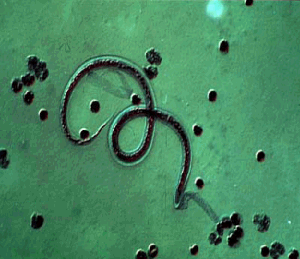 Lymphatic filariasis Lymphatic filariasis is a debilitating disease caused by nematode worms of the genera Wucheria and Brugia. Larval worms circulate in the bloodstream of infected persons, and adult worms live in the lymphatic vessels. Lymphatic filariasis is not life threatening, but it does cause discomfort, swelling of the limbs and genitals, damage to the kidneys and lymphatic system, impairment of the body's ability to fight infection, and general malaise. In addition, it causes immeasurable emotional and economic costs in terms of the disruption of family and community life. Humans contract filariasis when they are bitten repeatedly by mosquitoes infected with filarial worms. Over 70 species of mosquitoes in the genera Culex, Anopheles, Aedes, and Mansoniacan infect humans with the disease. Mosquitoes pick up the tiny, microfilarial form of the parasite when taking blood meals from infected humans. In the mosquito, the microfilariae develop within 7-21 days into members of the next stage of the parasite's life cycle, which are known as filariform larva. The filariform larva are infective to humans. When the larval worms move to a mosquito's mouth, and then the mosquito bites humans, the parasites can spread through a human community. Fortunately, however, many bites from infected mosquitoes are required before a person is infected with the disease. Once a human does pick up filariform larvae from mosquito bites, the larvae move to the lymphatic system, where they develop into adult worms. It usually takes 8-16 months after infection for symptoms of the disease to appear. The life span of adult worms is approximately seven years (microfilariae have a life span of from 3-36 months). The adults range in size from 2-50 cm in length. In the human, the adult worms mate and then the females produce millions of new microfilariae, which then circulate in the blood stream. Microfilariae circulating in the bloodstream can then be picked up by mosquitoes taking blood meals. In most endemic regions, microfilariae show peak abundance in the human bloodstream between 10 p.m. and 2 a.m., which corresponds with the time when Culex mosquitoes are most active. In some regions of the South Pacific, however, where the vectors of filariasis are active primarily in the daytime, microfilariae are most abundant during the day. These observations are consistent with the hypothesis that the microfilariae-abundance cycle in the bloodstream has evolved to maximize transmission to mosquitoes. [External Links]More Information from Boston College's websiteMore information from dhpe.orgMore information from wrongdiagnosis.com
|


 Posts
Posts

